EPFL scientists have used a snap-freezing method to show the structure of brain connections joining neurons together in human brains.
A new study shows size and shape of the dendritic spine determines information transmission
Normally, synaptic connections in the brain are located on dendritic spines, which are small, micro protrusions that extend from the neurons’ surface. Interestingly, the size and shape of one’s spine determine how well they pass signals between neurons. If the spine head is larger, then its synapse and receptor number and synaptic strength will be large. The spine neck separates the synapse on the spine from the dendrite and this often thin compartmentalizing signal creates electrical resistance between the dendrite and synapse. The morphology changes in the spine neck and head are witnessed after various activity forms such as LTP.
The details are vital when neuroscientists are looking at model brain circuits to understand the transmission of information between neurons and neuronal circuits in the brain. But, the small size and challenges in brain tissue preservation in its natural state have always left scientists concerned about what a dendritic spin looks like. The
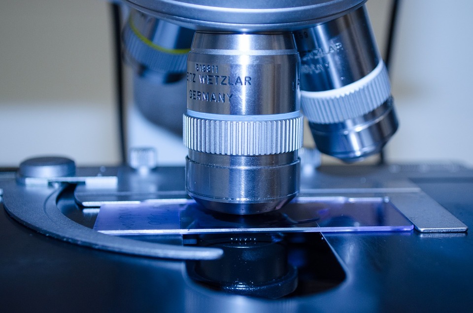
Now, EPFL School of Life Sciences scientists have used a snap-freezing method involving liquid nitrogen jets in combination with high pressure to preserve some small brain tissue pieces. The Carl Petersen and Graham Knott labs researchers also employed high-resolution 3D imaging using electron microscopes to show the true structure of the dendritic spine relative to past studies. Interestingly, the snap-freezing method indicated that dendritic spines had thinner necks.
Dendritic spines are electrical and chemical compartment
Most importantly, the finding validates throngs of past theoretical and functional data showing that dendritic spines are chemical and electrical compartments that are isolated from the neuron by a thin neck. The neck diameter variations have a significant impact on how a synapse influences the neuron.
Graham Knott said that besides revealing the true shape of the dendritic spine structures, their work also demonstrates the importance of electron microscopy and instantaneous freezing methods in getting a detailed view of cell and tissue architecture.


