
The Technical University of Munich (TUM) and Helmholtz Munich researchers have created an innovative method using AI and high-resolution optoacoustic imaging to measure microvascular changes in the skin, indicating the severity of diabetes-related small blood vessel alterations. Published in Nature Biomedical Engineering, this advancement enables more precise disease assessment.
Researchers identify 32 skin changes predicting possibility of diabetes
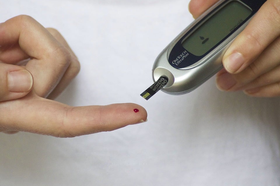
Optoacoustic imaging is a technique that utilizes light pulses to create ultrasound waves within tissue. These waves are detected by sensors and translated into images. The signals are generated by the movement of tissue surrounding molecules that absorb light, such as hemoglobin. Due to the high concentration of hemoglobin in blood vessels, optoacoustic imaging can provide highly detailed and distinctive images of vessels that cannot be obtained through other non-invasive methods.
It is important to note that optoacoustics, principles have been existing for over a century and are now finding their way to medical applications. Vasilis Ntziachristos, TUM Professor, developed optoacoustic imaging methods, including RSOM (raster-scan optoacoustic mesoscopy), at the Institute of Biological and Medical Imaging and Helmholtz Munich’s Bioengineering Center.
Researchers utilized RSOM to examine diabetes effects on human skin, studying 75 diabetic legs and a control group. Applying an AI algorithm to RSOM images, they identified 32 noteworthy changes in skin microvasculature, encompassing vessel branch number and diameter, advancing understanding of diabetes-related alterations.
RSOM a faster non-invasive technique of measuring vascular changes
RSOM enables rapid, non-invasive measurement of vascular changes in diabetic patients, offering advantages over biopsy-based methods. Unlike invasive biopsies, RSOM provides real-time observations without blood vessel deformation. According to lead clinician Angelos Karlas the procedure takes less than a minute, doesn’t involve radiation or contrast agents, and surpasses other optical methods in depth and detail.
A single RSOM measurement allows simultaneous data collection from different skin depths, revealing that diabetes impacts vessels in distinct skin layers. The dermal layer shows reduced vessel numbers, while the epidermal layer exhibits an increase. Combining 32 skin characteristics provides a comprehensive assessment of diabetes severity, establishing a novel link between skin blood vessels and disease severity.